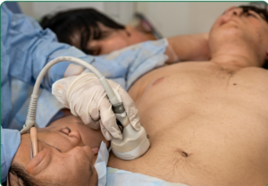
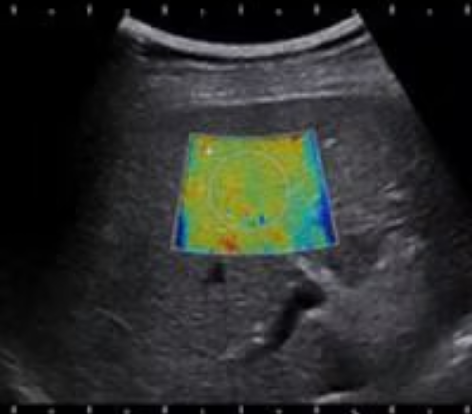
Meenakshi Radiology

Phone: +91 82485 90535
Address: Near New Bus stand, Tanjore

Usg Elastography

We welcome 

Our first batch of DNB Radiology students in Meenakshi hospital, Tanjore

Non-invasive Assessment of Tissue Stiffness

What is Elastography?

Elastography is an ultrasound-based imaging technique that measures tissue stiffness (elasticity). Stiffer tissues (e.g., fibrosis, tumors) show less deformation compared to softer tissues.


Diagram Labels

Indications

Liver

Focal Lesions

Other Indications

Musculoskeletal

STEPS TO PERFORM LIVER SHEAR WAVE ELASTOGRAPHY

USG-E

01.B-mode image

1

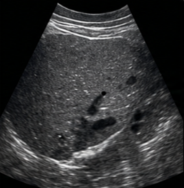
Obtain good quality 2D image

Jan

02. Place ROI

2

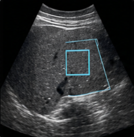
1.0-2.0 cm below glisson capsule, avoid vessels

Jan

03.Activate SW Elastography

3

Keep patient in end-expiration

Jan

04. Check quality criteria

4

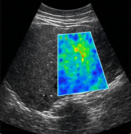
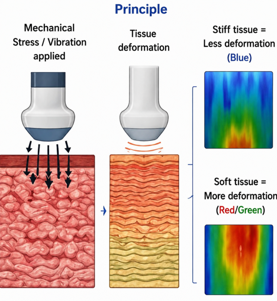
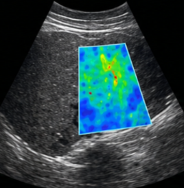
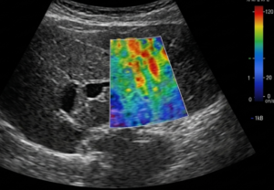
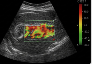
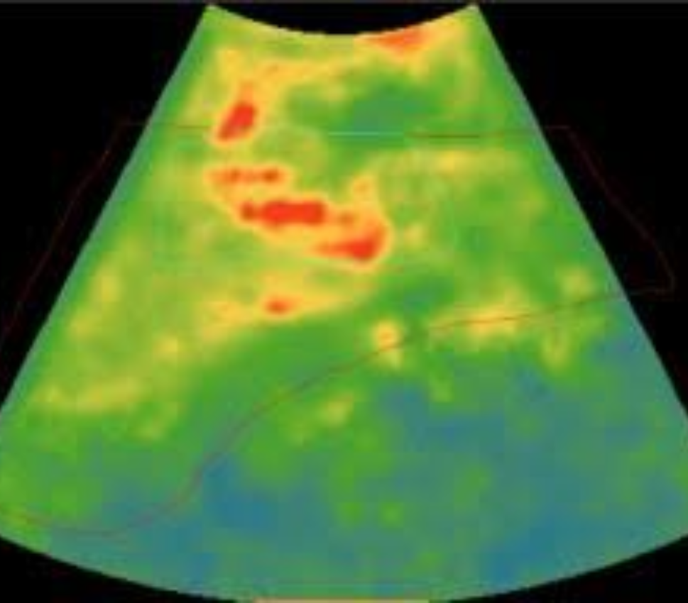
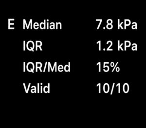
Good map, valid meas, ≥ 60%

Jan

05.Record measurements

5

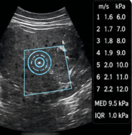
Take ≥ 10 valid measurements and record median (kPa)
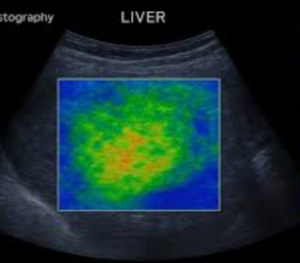
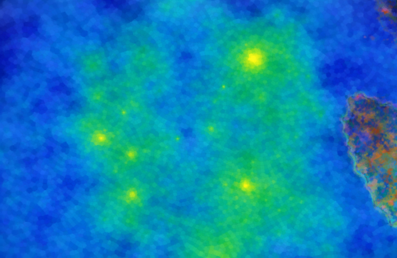
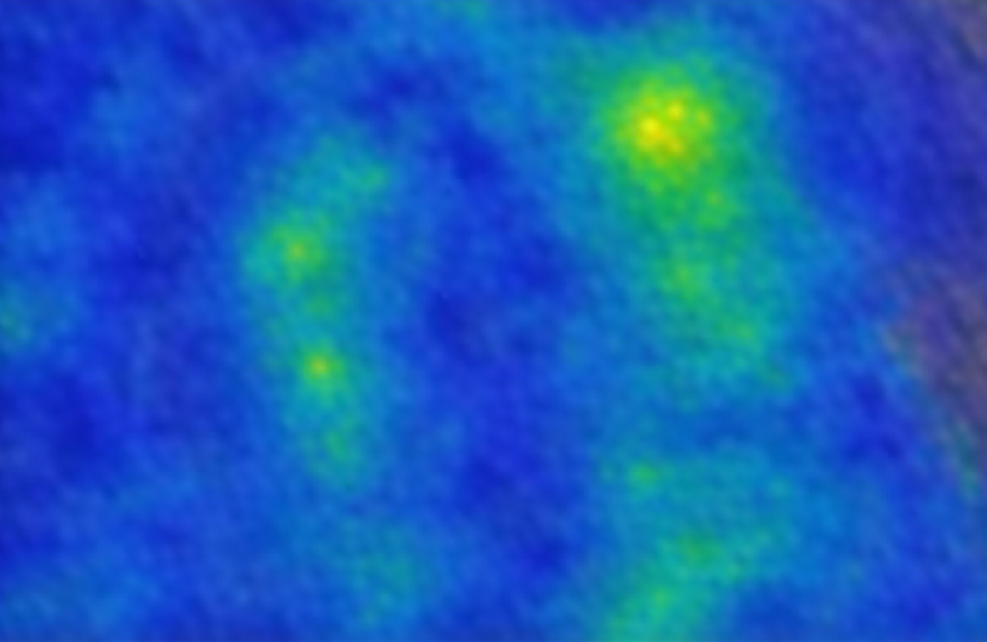
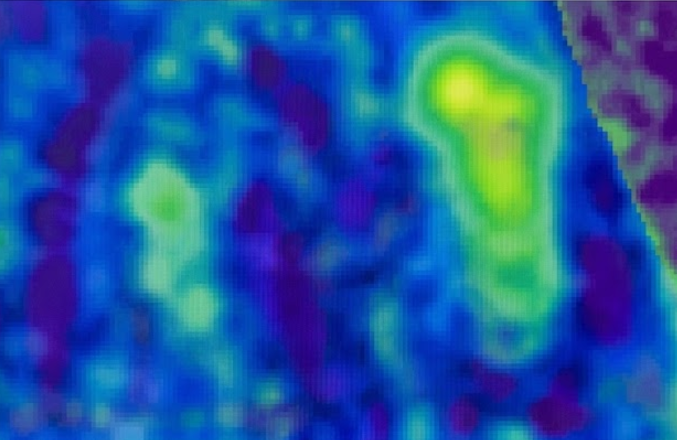
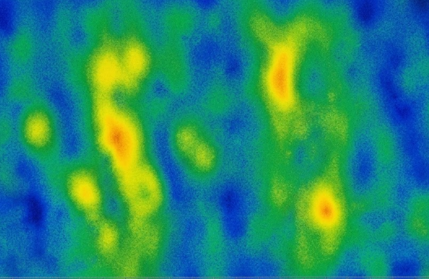
Jan

TECHNIQUE

Patient preparation
Patient position
Transducer
ROI Placement
Acquisition
Measurement

TYPES OF ELASTOGRAPHY

STRAIN ELASTOGRAPHY (QUALITATIVE/SEMI-QUANTITATIVE

SHEAR WAVE ELASTOGRAPHY (QUANTITATIVE)

ADVANTAGES

Normal vs Pathological Findings (Liver – Shear Wave)

Fibrosis Stage (Metavir) Description Shear Wave Velocity (kPa) Elastography Image (Color Map)
F0 – F1 No / Mild fibrosis ≤ 7.0 F0-F1
F2 Moderate fibrosis 7.1 – 9.5 F2
F3 Advanced fibrosis 9.6 – 12.5 F3
F4 Cirrhosis > 12.5 F4
* Cut-off values vary with etiology and equipment.

Key Points

Measures tissue stiffness (Elasticity)
Helps in staging fibrosis and characterizing lesions
Shear Wave Elastography provides quantitative values in kPa
Operator skill and proper technique are essential
Integrate findings with clinical and other investigations

Contraindications

Important Advice

Note