Meenakshi Radiology

Phone: +91 82485 90535
Address: Near New Bus stand, Tanjore

Slow Flow Venous Malformation – Embolisation

We welcome 

Our first batch of DNB Radiology students in Meenakshi hospital, Tanjore

Minimally invasive image-guided therapy for symptomatic venous malformations

What is a Slow Flow Venous Malformation?

Venous malformations (VMs) are congenital, abnormal, dilated venous channels with slow blood flow. They can cause progressive swelling, pain, disfigurement, functional impairment and complications like thrombosis, bleeding and ulceration.


Indications

Embolisation Technique – Step by Step

SFVME

01.Vascular Access

1

Percutaneous access (usually femoral vein) under ultrasound guidance.

Jan

02.Diagnostic Venography

2

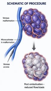
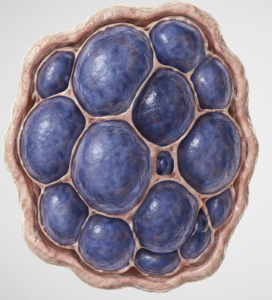
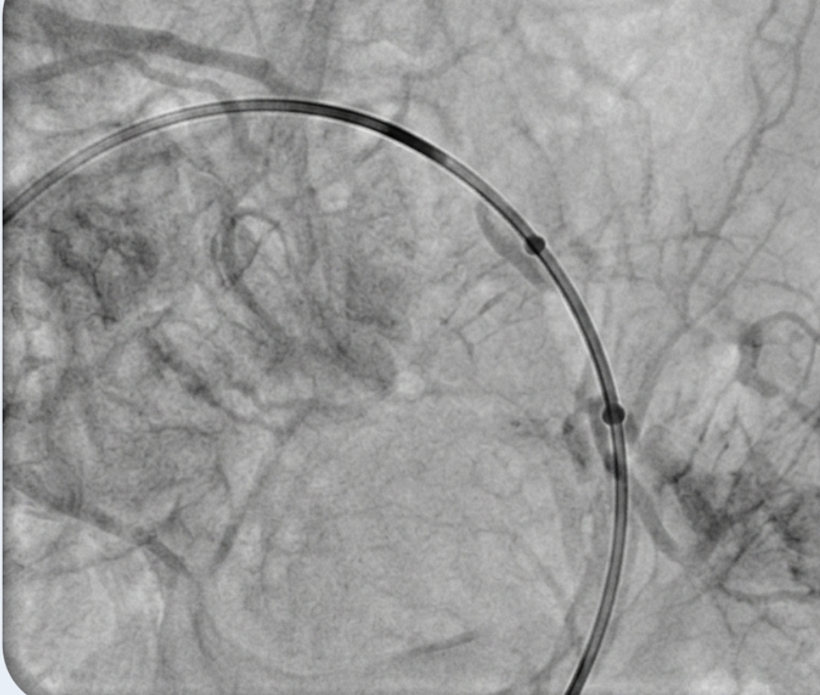
Catheter navigation into the malformation and venography to define anatomy, extent and drainage.

Jan

03. Microcatheter Placement

3

Superselective catheterisation of the malformation nidus/venous lakes.

Jan

04.Embolic Agent Injection

4

Slow, controlled injection of sclerosant/embolic agent until stasis is achieved.

Jan

05.Post Embolisation Venography

5

To assess reduction in flow and adequate penetration.

Jan

COMMON EMBOLIC AGENTS USED

ADVANTAGES

Minimally Invasive

Repeatable & Safe
Reduces Lesion Size
Symptom Relief

Adjunct To Surgery

Contraindications

Potential Complications

IMPORTANT POINTS

Important Advice