Meenakshi Radiology

Phone: +91 82485 90535
Address: Near New Bus stand, Tanjore

CT-Guided Biopsies

We welcome 

Our first batch of DNB Radiology students in Meenakshi hospital, Tanjore

Minimally Invasive | Image Guided | Accurate Diagnosis

What Is Ct-Guided Biopsy?

A minimally invasive image-guided procedure in which CT imaging is used to accurately place a needle into a lesion to obtain tissue for histopathology or cytology.

Indications

Lung Mass

Mediastinal Tumors

Retroperitoneal Nodes

Procedure Technique: Step By Step

CT-GB

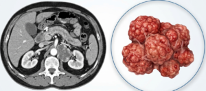
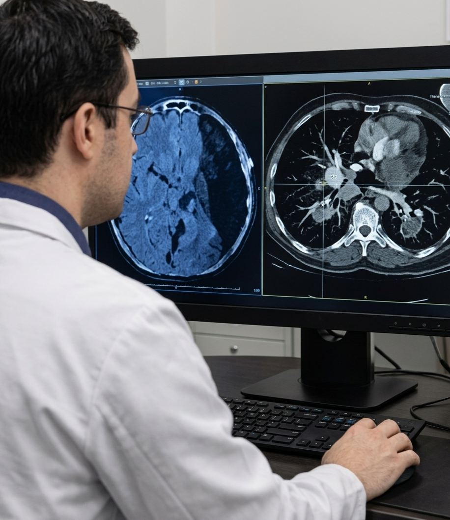
01.Pre-procedure Planning

1

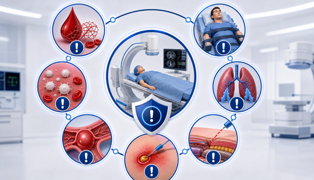
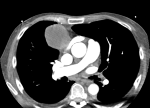
Review clinical details, previous imaging. Choose safest and shortest needle path. Check coagulation profile & consent.

Jan

02.Patient Positioning

2

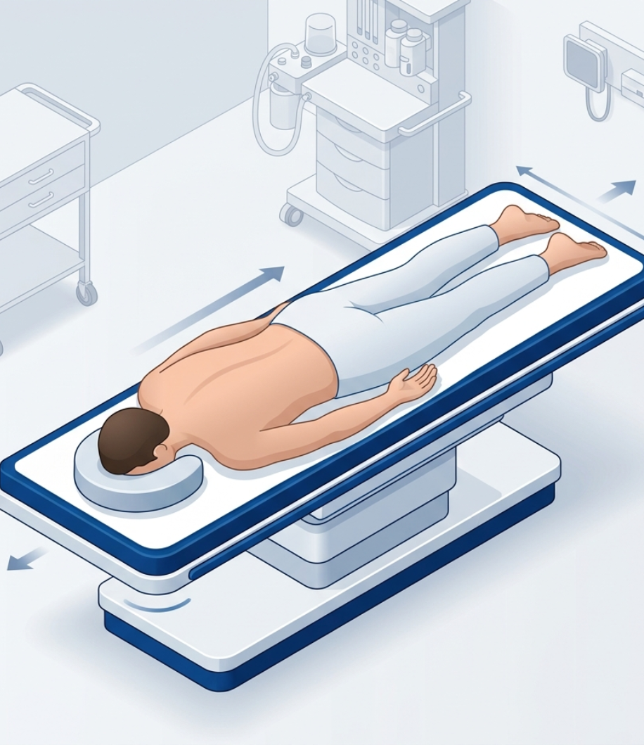
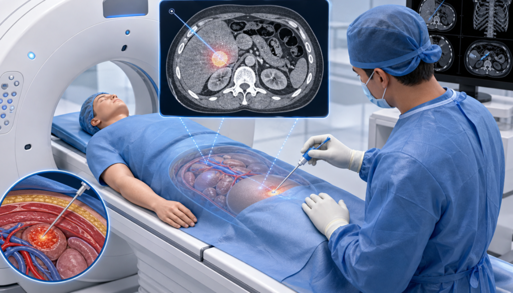
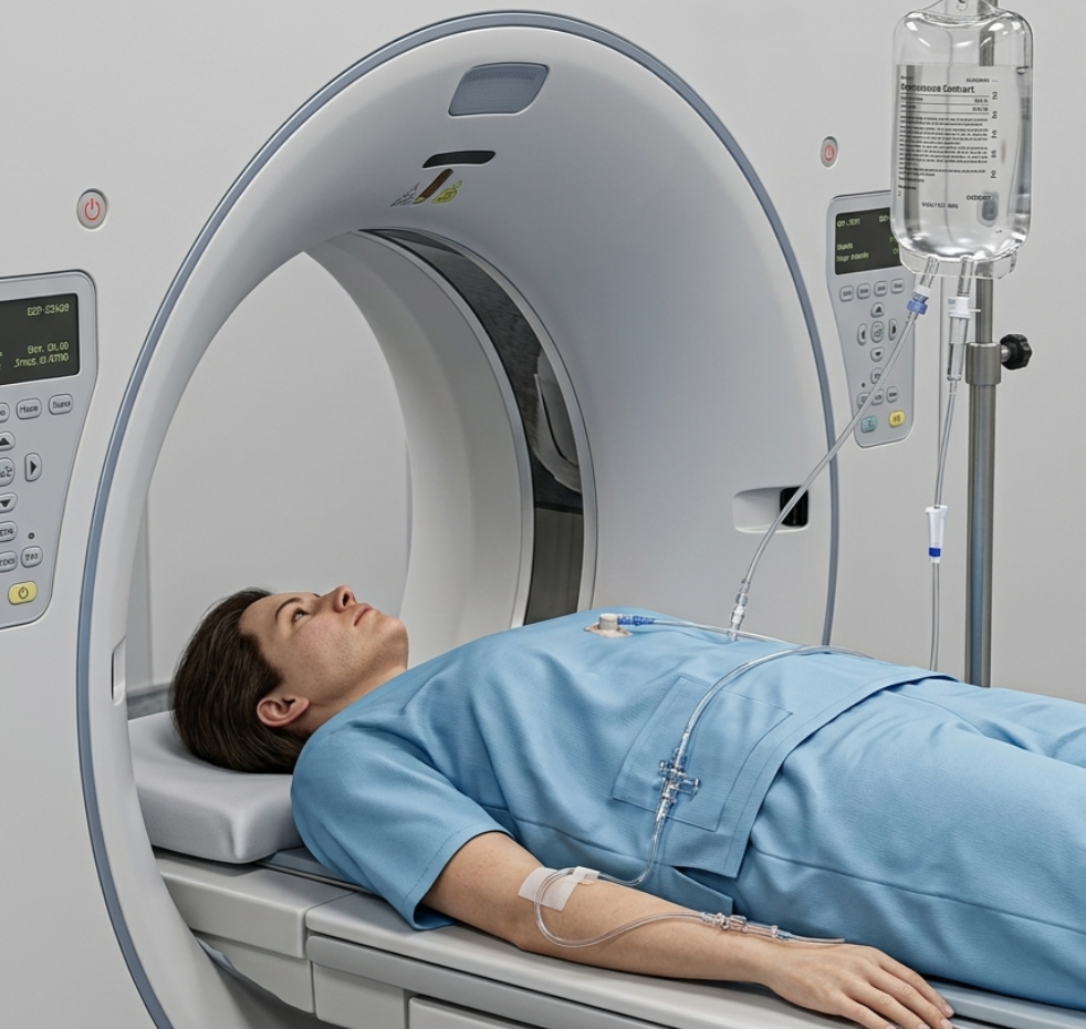
Position patient (prone/supine/lateral) depending on lesion location.

Jan

03.Sterile Preparation & Local Anaesthesia

3

Aseptic cleaning and draping. Local anaesthesia to skin and deeper tissues.

Jan

04.Needle Insertion under CT Guidance

4

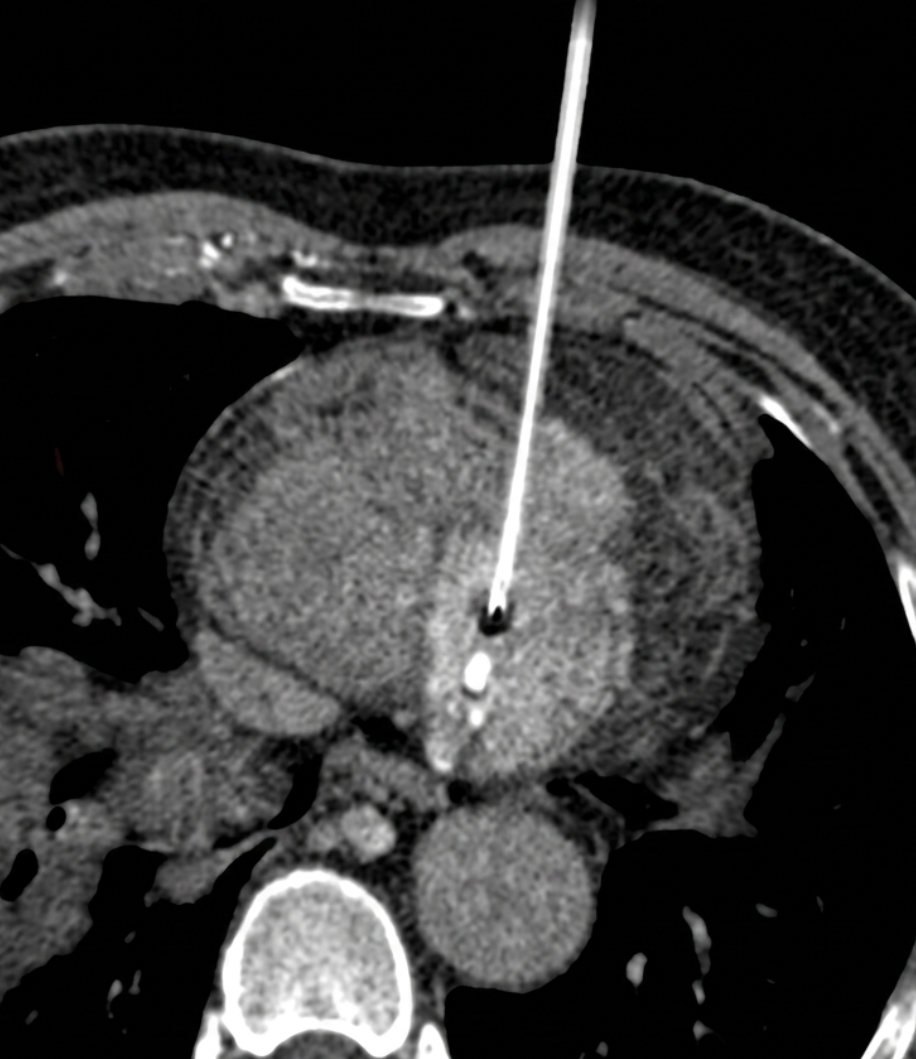
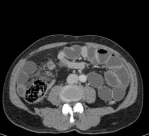
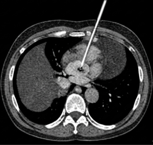
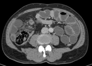
Stepwise needle advancement with intermittent CT scans. Coaxial technique commonly used.

Jan

05.Tissue Sampling

5

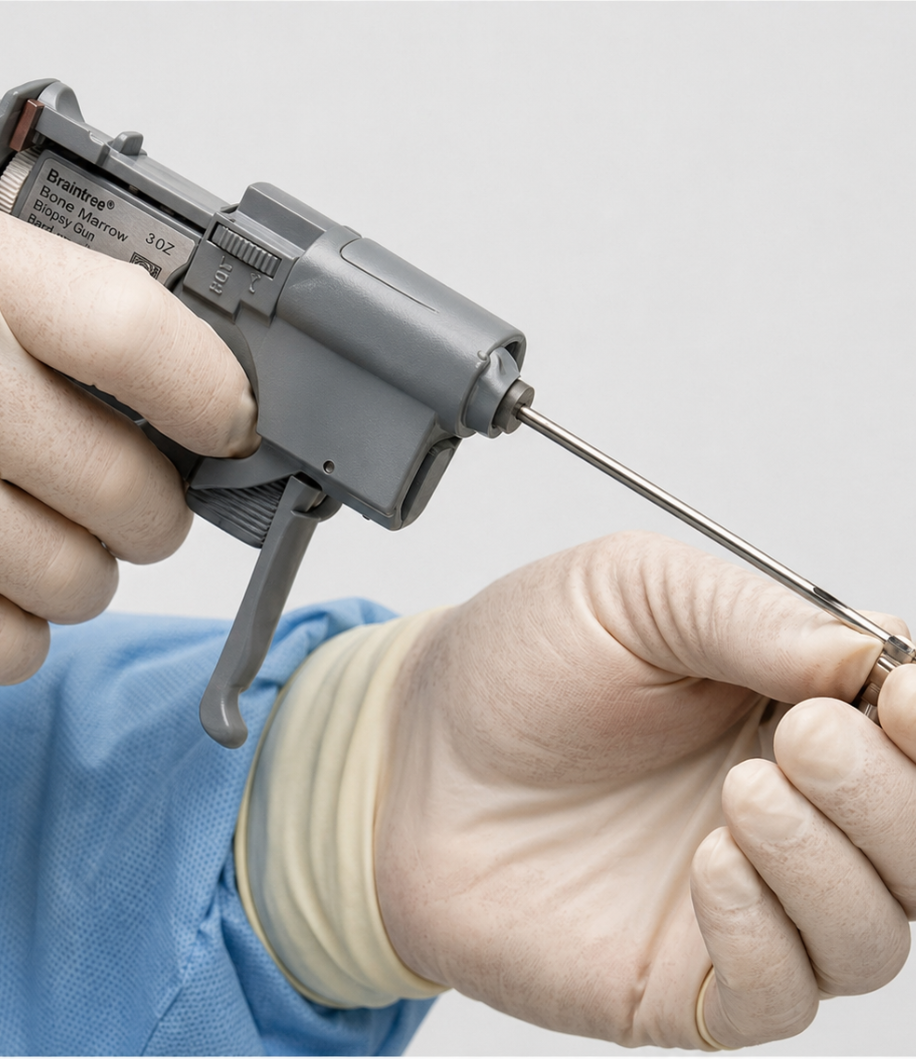
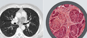
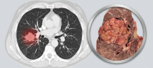
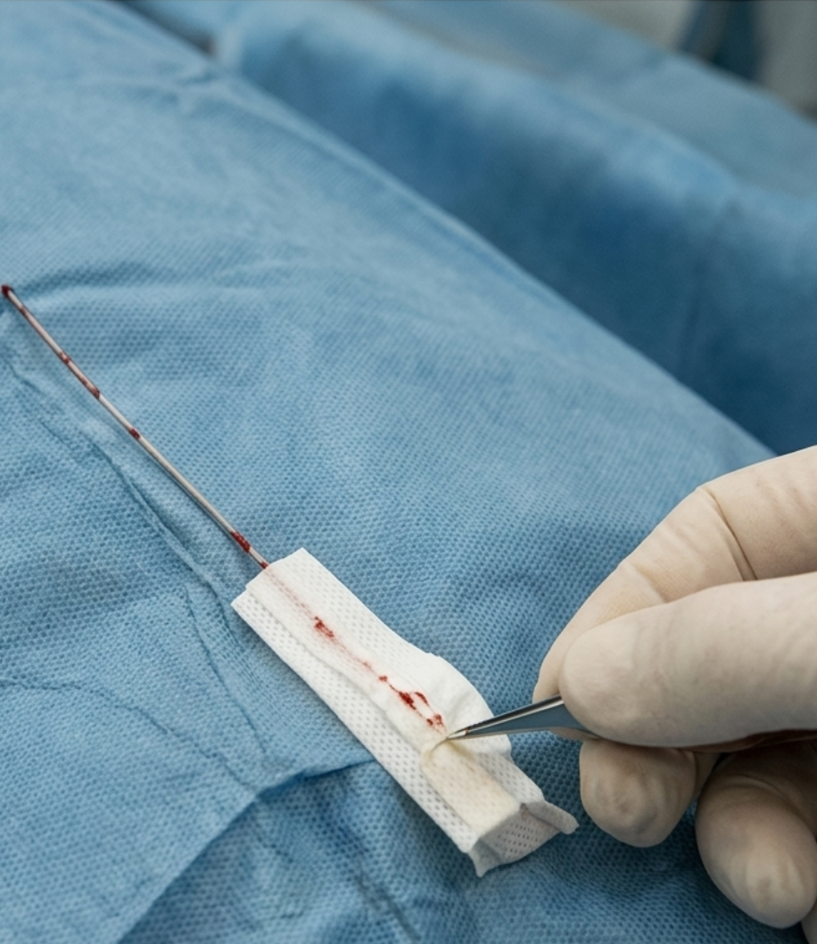
Obtain FNAC and/or core tissue using biopsy needle or automated biopsy gun.

Jan

06.Post-procedure Scan

6

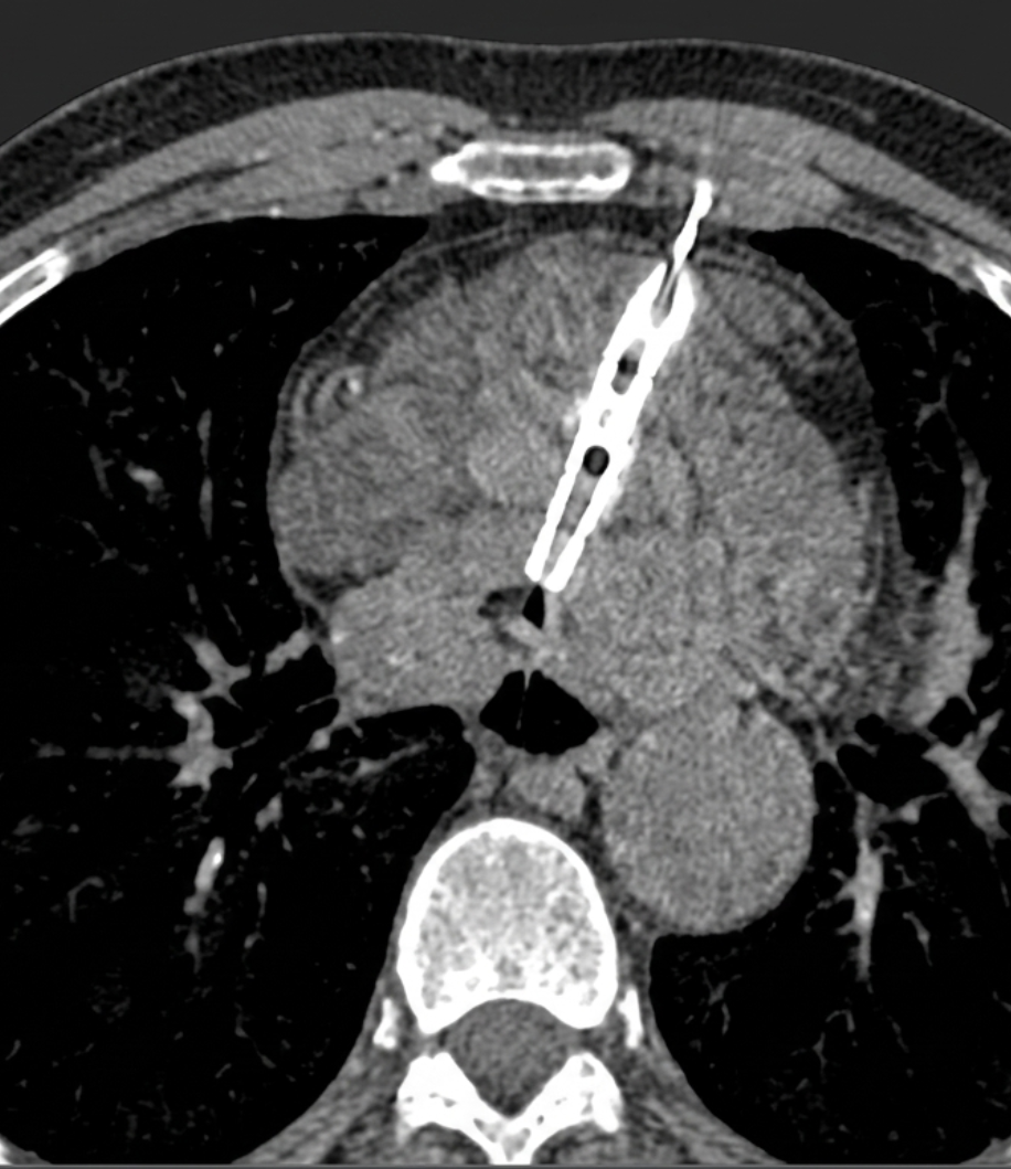
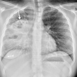
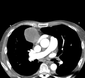
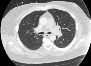
Immediate CT scan to check for complications (pneumothorax, bleeding, etc.).

Jan

07. Observation

7

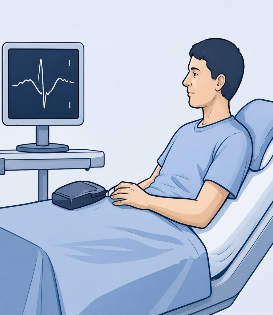
Observe patient for 2-4 hours (longer for lung biopsy). Monitor vitals and symptoms.

Jan

Contraindications

ADVANTAGES

Minimally invasive

High diagnostic accuracy

Real-time precise targeting

Avoids open surgical biopsy

Wide range of accessible lesions

Short hospital stay / day care procedure

Cost-effective with good safety profile

Potential Complications

Lung Biopsy

Mediastinal Biopsy

Retroperitoneal Biopsy

Key Points

Examples Of Lesions

Lung Mass
Mediastinal Tumor
Retroperitoneal Node

Important Advice

Note