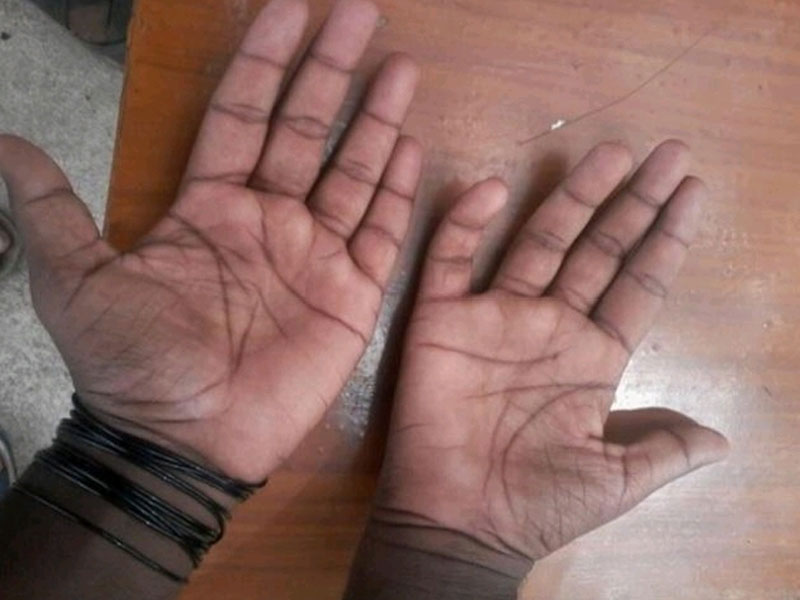
A 19-year-old male patient presented with unilateral upper limb weakness for the last eight months. Neurological examination revealed weakness and muscle wasting in right forearm and hand (Fig. 1), without sensory disturbance.
No sensory or motor deficit elicited in left upper limb or in bilateral lower limbs.
Final Diagnosis: Juvenile asymmetric segmental spinal muscular atrophy (JASSMA) or Hirayama disease.
Imaging Findings:
Cervical MRI plays an important role in the diagnosis of this rare disease. Imaging in our case is done with 1.5 T MRI (Philips Multiva). Imaging protocol includes T1-and T2-weighted imaging in both axial and sagittal planes and in both neutral and flexion positions. MR imaging in neutral position demonstrates the subtle focal cervical spinal cord atrophy (Fig. 2 & 3), especially in the lower cervical region. Flexion of neck is achieved by placing the custom built positioning sponges under head. Imaging in this position, additionally elicits the anterior displacement of dura with resulting widening of the posterior epidural space (Fig. 4) and more pronounced cord thinning, which was not obvious in neutral position (Fig. 5).